Nature | News Feature
Sharing
Crystallography: Atomic secrets
100 years of crystallography.
Article tools
In 1914, German scientist Max von Laue won the Nobel Prize in Physics for discovering how crystals can diffract X-rays: a phenomenon that led to the science of X-ray crystallography. Since then, researchers have used diffraction to work out the crystalline structures of increasingly complex molecules, from simple minerals to high-tech materials such as graphene and biological structures, including viruses. With improvements in technology, the pace of discovery has accelerated: tens of thousands of new structures are now imaged every year. The resolution of crystallographic images of proteins passed a critical threshold for discriminating single atoms in the 1990s, and newer X-ray sources promise images of challenging proteins that are hard or impossible to grow into large crystals.
Birth of an idea
 SPL
SPL
Von Laue hit on the idea that when X-rays passed through a crystal, they would scatter off the atoms in the sample and then interfere with each other like waves passing through a breach in a shore wall. In some places, the waves would add to each other; in others, cancel each other out. The resulting diffraction pattern could be used to back-calculate the location of the atoms that scattered the original X-rays. Von Laue and his colleagues proved his theory in 1912 with a sample of copper sulphate.
Going up
The Worldwide Protein Data Bank has been collecting resolved structures of proteins since 1971, and now holds nearly 100,000 entries. Other databanks, including the Crystallography Open Database (COD), include structures of everything from minerals to metals and small biological molecules. The COD is now adding instructions into its database for how to print three-dimensional models of some structures.
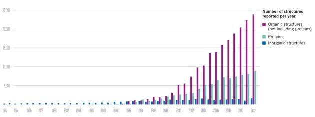 Source: Worldwide Protein Data Bank/ Crystallography Open Database
Getting clearer
Better techniques for both imaging and interpreting data have allowed researchers see finer details in some structures and tackle ever more complicated molecules.
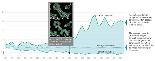 Images: Bernhard Rupp/Garland Science/Taylor & Francis Graph: H. M. Berman Protein Sci. 21, 1587–1596 (2012), with updates from Worldwide Protein Data Bank
100 years of crystallography
1913: Diamond
Diffraction image allowed researchers to confirm the tetrahedral structure of carbon atoms in this famous crystal.
1923: Hexamethylenetetramine
The first organic molecule to be imaged, chosen because of its simple cubic symmetry. It proved that molecules, not just atoms, can make up the repeating elements of a crystal.
 Am. Chem. Soc.
Hexamethylenetetramine in 3D
Courtesy: James Garnett and Jonathan Taylor
1925: Quartz
The determination of the structure of silicate minerals was fundamental to the field of mineraology.
1952: DNA
Rosalind Franklin’s X-ray image of DNA, known as photo 51, helped James Watson and Francis Crick to create their famous model of the double helix. An atomic-resolution image of the structure proposed in 1953 was not taken until 1980.
 King’s College London
DNA in 3D
Courtesy: James Garnett and Jonathan Taylor
1958: Myoglobin
The irregular folds seen in the structure of the first imaged protein were a huge surprise.

Myoglobin in 3D
Courtesy: James Garnett and Jonathan Taylor
1965: Lysozyme
The first enzyme to be imaged, sourced from hen egg whites.

Lysozyme in 3D
Courtesy: James Garnett and Jonathan Taylor
1970: Synchrotron
A study of insect muscle at the German Electron Synchrotron (DESY) in Hamburg was the first to use X-rays generated by a synchrotron. The use of these machines caused a boom in crystallography studies.
1978: Tomato bushy stunt virus
First atomic-scale image of a complete virus: in this case, a plant virus. It revealed structural rules that were found to hold true in human pathogens a few years later.

Tomato bushy stunt virus in 3D
Courtesy: James Garnett and Jonathan Taylor
1984: Quasicrystals
The first crystals were identified with atomic arrangements that do not repeat exactly, defying general wisdom about crystals.
 US Dept of Energy/AFP/Getty
2000: Ribosome
The molecular machine that assembles proteins from instructions encoded in DNA.
 V. Ramakrishnan & D. E. Brodersen/Medical Research Council
Ribosome in 3D
Courtesy: Said Sannuga and V. Ramakrishnan, MRC Laboratory of Molecular Biology
2009: X-ray free-electron laser
The Linac Coherent Light Source at the SLAC National Accelerator Laboratory in Menlo Park, California, went into operation, opening up a new world of imaging possibilities (see page 604).
2013: HIV trimer
An X-ray crystallographic image of the hook that HIV uses to bind to human cells helped to resolve a debate about what this important protein looks like.
 AAAS
AAAS
HIV trimer in 3D
Courtesy: James Garnett and Jonathan Taylor
The future
The ‘most wanted’ list of proteins that remain to be imaged includes the massive spliceosome, which helps to organize and edit messenger RNA, and the even larger nuclear-pore complex, which serves as a nucleus’s gatekeeper.
These structures can contain hundreds of proteins, making them hard to crystallize or keep still for an image.
One strategy is to crystallize bits of these structures and piece them together like a jigsaw; the use of X-ray free-electron lasers should also help.
- Nature
- 505,
- 602–603
- (30 January 2014)
- doi:10.1038/505602a
For the best commenting experience, please login or register as a user and agree to our Community Guidelines. You will be re-directed back to this page where you will see comments updating in real-time and have the ability to recommend comments to other users.
Leave a Reply
You must be logged in to post a comment.